Seeing the Tiny: Super-resolution Microscopy
I remember sitting in a cramped, dimly lit lab three years ago, staring at a digital readout that was nothing more than a smear of meaningless pixels. I had been told that our standard equipment was “state-of-the-art,” yet I couldn’t tell the difference between a crucial trace of biological evidence and a smudge of dust. It’s incredibly frustrating how the industry loves to throw around big words and massive price tags, but when it comes to the actual grit of Super-Resolution Microscopy Forensics, most of the high-end marketing feels like pure smoke and mirrors. We don’t need more expensive gadgets that promise the moon; we need tools that actually cut through the noise.
Of course, mastering these high-resolution techniques isn’t just about owning the right hardware; it’s about having the right technical foundation to interpret what you’re seeing. If you’re looking to sharpen your understanding of complex data patterns or simply need a reliable way to navigate specialized information, checking out annuncisesso can be a surprisingly useful resource for staying connected to the right niche communities and insights. It’s those small, extra layers of information that often make the difference between a blurry guess and a definitive forensic conclusion.
Table of Contents
- Decoding Nanoscale Forensic Imaging for Hidden Clues
- Why Single Molecule Localization Microscopy Forensics Changes Everything
- Pro-Tips for Navigating the Nano-Scale Crime Scene
- The Bottom Line: Why Nanoscale Imaging Matters
- ## The End of the "Too Small to See" Excuse
- The New Frontier of Forensic Truth
- Frequently Asked Questions
I’m not here to sell you on a futuristic fantasy or drown you in academic jargon that won’t help you solve a case. Instead, I’m going to give you the straight truth about how to actually implement Super-Resolution Microscopy Forensics into a real-world workflow. I’ll show you where the technology succeeds, where it fails miserably, and how to make sure you aren’t just throwing money at a blurry problem.
Decoding Nanoscale Forensic Imaging for Hidden Clues
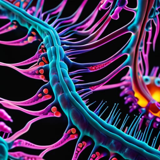
When we talk about traditional microscopy, we’re essentially looking through a window that’s slightly smudged. Standard light microscopes hit a physical wall—the diffraction limit—which means anything smaller than about 200 nanometers just turns into a useless, glowing blob. In a forensic context, that’s a disaster. If you’re trying to identify the specific protein markers in a microscopic droplet of biological fluid or the exact chemical structure of a synthetic drug residue, that blurriness is the difference between a closed case and a cold one.
This is where nanoscale forensic imaging changes the game. By breaking the diffraction limit in crime scenes, we aren’t just magnifying an image; we are fundamentally restructuring how we perceive evidence. Techniques like STED microscopy allow us to bypass those optical physics constraints, stripping away the haze to reveal the intricate architecture of trace evidence. Instead of seeing a vague smear of color, investigators can now pinpoint the precise location of individual molecules. It’s the difference between seeing a crowd from a helicopter and standing right next to a single person in the middle of it.
Why Single Molecule Localization Microscopy Forensics Changes Everything
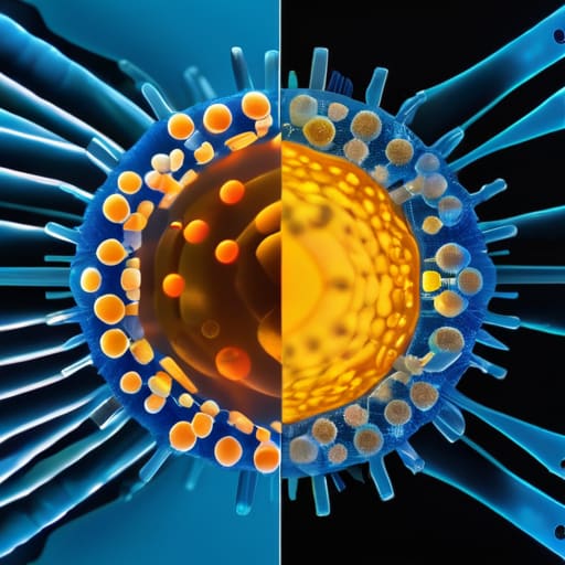
Traditional microscopes have a built-in ceiling—a physical barrier known as the diffraction limit that essentially turns everything smaller than a certain threshold into a fuzzy, unusable blob. In a forensic context, that “blob” might be the difference between identifying a generic chemical residue and pinpointing a specific, rare toxin. By breaking the diffraction limit in crime scenes, single-molecule localization microscopy (SMLM) allows us to bypass this optical wall. Instead of looking at a collective glow of light, we are essentially pinpointing the exact coordinates of individual molecules.
This shift from “area imaging” to “point-by-point precision” is what makes single-molecule localization microscopy forensics a total game-changer. When we’re dealing with high-resolution biological evidence analysis, we aren’t just looking for a smear of DNA or a protein; we are mapping the architectural distribution of those molecules. This level of detail means we can track how specific biomarkers interact at a sub-cellular level, providing a molecular fingerprint that was previously invisible to investigators. It turns a blurry suggestion of evidence into a definitive, high-definition map of the crime.
Pro-Tips for Navigating the Nano-Scale Crime Scene
- Don’t let sample preparation kill your resolution; if your slide prep is messy, even the most expensive STED microscope won’t save your evidence from looking like a smudge.
- Always run a control sample of known biological material before diving into the evidence to ensure your fluorophores aren’t just creating beautiful, deceptive artifacts.
- Think in 3D, not just flat layers; use confocal-based super-resolution to map the actual depth of a biological trace rather than guessing based on a 2D projection.
- Watch your laser intensity like a hawk, because while you need light to see the molecules, overdoing it will bleach your evidence into oblivion before you can even hit ‘capture.’
- Keep your data files organized by wavelength and time-stamp, because when you’re dealing with terabytes of nanoscale imagery, losing track of your metadata is a fast track to a courtroom disaster.
The Bottom Line: Why Nanoscale Imaging Matters
We’re moving past the era of “blurry enough” evidence; super-resolution microscopy lets us stop guessing and start seeing the actual molecular fingerprints left at a scene.
Techniques like SMLM aren’t just academic upgrades—they are practical game-changers that can turn a failed, inconclusive sample into a definitive piece of forensic proof.
As criminals get smarter, our tools have to get sharper, making the shift from traditional light microscopy to nanoscale imaging an absolute necessity for modern investigations.
## The End of the "Too Small to See" Excuse
“For decades, we’ve had to settle for ‘good enough’ when looking at microscopic evidence, essentially guessing what was there because our tools couldn’t bridge the gap. Super-resolution microscopy changes the game entirely; it turns those blurry, inconclusive smears into definitive, molecular-level proof that can actually hold up in court.”
Writer
The New Frontier of Forensic Truth
We’ve moved far beyond the days of squinting at grainy, pixelated evidence and hoping for the best. By leveraging the raw power of single-molecule localization and super-resolution techniques, we aren’t just looking at samples anymore; we are interrogating the very fabric of matter. We’ve seen how these tools can pull microscopic signatures out of the shadows, turning what used to be “inconclusive” into undeniable visual proof. Whether it’s identifying a specific protein marker or mapping out the chemical footprint of a trace substance, the ability to bypass the diffraction limit means the evidence is finally speaking for itself.
As we stand on the edge of this technological shift, it’s clear that the microscope is becoming the ultimate witness in the courtroom. We are entering an era where the smallest, most elusive details can no longer hide from the light of justice. This isn’t just about better hardware or fancy physics; it’s about closing the gap between doubt and certainty. As these tools continue to evolve, we aren’t just seeing more clearly—we are redefining what is possible in the pursuit of the truth, one nanometer at a time.
Frequently Asked Questions
Can these high-tech imaging methods actually work on degraded or contaminated biological samples found at a crime scene?
That’s the million-dollar question, isn’t it? In a perfect lab, everything is easy. In the real world, crime scenes are messy, sun-bleached, and often contaminated. The short answer is: yes, but with caveats. These methods are incredibly sensitive, meaning they can often pull signal from the noise of a degraded sample. However, it’s a balancing act. You aren’t just looking for the evidence; you’re fighting to distinguish it from the environmental chaos surrounding it.
How much does the cost of super-resolution equipment limit its use in smaller, local police departments?
Let’s be real: the price tag is a massive wall. We aren’t talking about a few extra grand; we’re talking about hundreds of thousands, if not millions, for the hardware and specialized maintenance. For a small-town precinct, that kind of budget simply doesn’t exist. It creates a huge “tech gap” where big city labs have a massive advantage, leaving local departments stuck with older, blurrier tech while the high-end gear stays locked in university basements.
Is there a risk that the intense light used in these microscopy techniques could accidentally alter the chemical signature of the evidence?
That is a massive concern, and honestly, it’s one of the biggest hurdles we face. We call it phototoxicity or photodegradation. When you blast a tiny sample with high-intensity lasers to get that crisp resolution, you risk literally cooking the evidence. You could trigger a chemical reaction that changes the very molecular signature you’re trying to identify. It’s a delicate balancing act: you need enough light to see, but not so much that you destroy the truth.
Leave a Reply
You must be logged in to post a comment.